
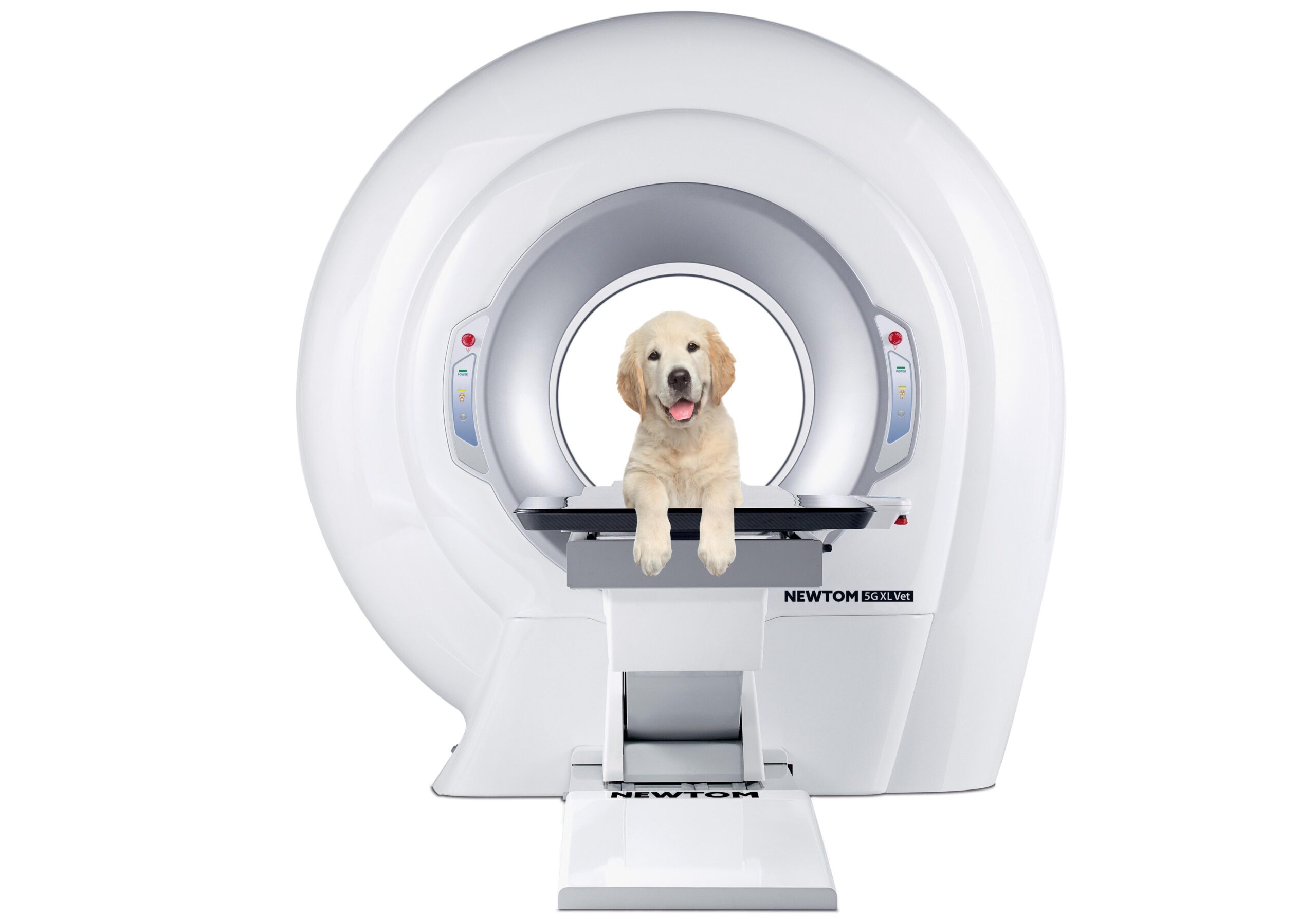

NewTom 5G XL VET – veterinary 3D computed tomography
NewTom 5G XL VET is a CBCT-based veterinary computed tomography system designed to obtain detailed 2D and 3D images. The system is suitable for a variety of veterinary examinations, including orthopedic diagnostics, upper respiratory tract, ear, dental and jaw pathology examinations.
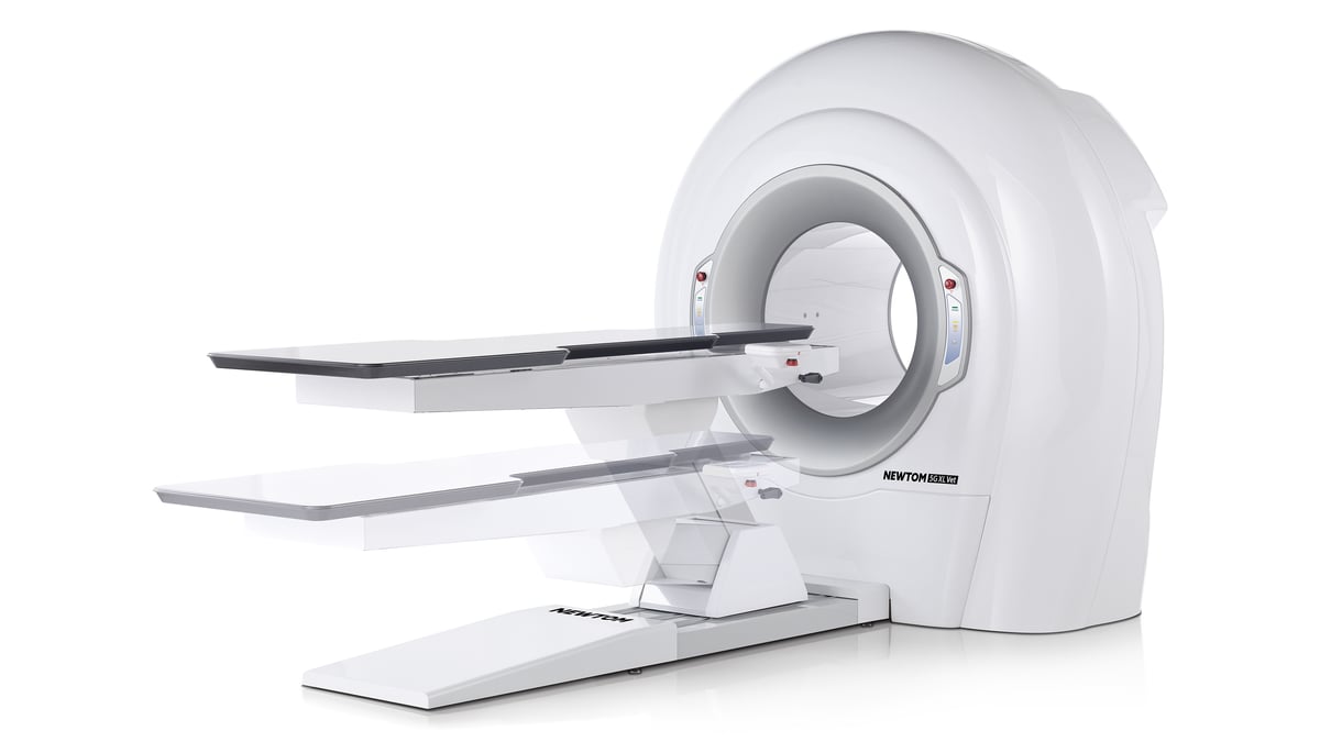
The open gantry and motorized carbon fiber patient table allow for comfortable positioning of the animal. A wide selection of fields of view (FOV) allows for customized imaging for different anatomical areas. SafeBeam™ technology automatically calculates the required radiation dose based on the patient’s anatomical characteristics, and the ECO Scan mode allows for shorter exposure times.
Main advantages
- High-quality 2D and 3D images: CBCT technology provides detailed images of anatomical structures.
- Wide range of research fields: from 6 × 6 cm to 21 × 19 cm, expandable to 21 × 28 cm using the eXtra FOV function.
- High definition video: selectable voxel size of 100-150 µm in HiRes mode.
- Motorized patient table: made of carbon fiber, allows you to remotely adjust the animal's position.
- SafeBeam™ technology: automatically adjusts the radiation dose according to the patient's anatomical characteristics.
- ECO Scan mode: During standard examinations, the duration of X-ray exposure can be reduced to 0.9 seconds.
- Ray2D function: allows for low-dose 2D X-ray examinations.
- CineX feature: enables the analysis of moving anatomical structures using sequences of X-ray images.
Diagnostic visualization
With the 5G XL VET, Newtom is adapting CBCT technology to new veterinary applications. Extremely high-quality 2D and 3D images, a wide range of fields of view (FOV) and specialized software.
- Dislocation of the lower jaw joint


- Lumbar disc herniation


- Nasal tumor


- Patellar dislocation before and after surgery


Technical specifications
| Parameter | Meaning |
|---|---|
| Technology | CBCT |
| Types of research | 2D and 3D |
| 3D scanning duration | 18-36 seconds |
| 3D exposure time | 0.9-9.0 seconds |
| Voxel size, standard mode | 200-300 µm |
| Voxel size, HiRes mode | 100-150 µm |
| Minimum FOV | 6 × 6 cm |
| Main FOV | 21 × 19 cm |
| Maximum FOV with eXtra FOV | 21 × 28 cm |
| X-ray tube focal spot size | 0.3mm |
| Image sensor | Amorphous silicon flat panel detector |
| Grayscale levels | 16 bit |
| Patient positioning | Lying down |
| Reconstruction duration | Less than 1 minute |
| Software | NewTom NNT |

Reviews
There are no reviews yet.