


3D scanning and features
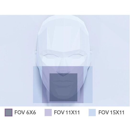
Personalization and precision with adaptive fields of view (FOV) ensure personalized imaging and maximum diagnostic quality: NewTom VG-One offers a wide choice of fields of view (FOV), enabling 3D scanning that targets specific anatomical areas and limits radiation to the area of clinical relevance.
Adaptation to the patient
The FOV function ensures that the field of view matches the patient and diagnostic requirements, allowing sectors to be scanned and the dose to be focused on the relevant details.
Configuration
VG One is designed to integrate seamlessly into any dental practice. Its configuration options allow it to be adapted to all diagnostic and clinical requirements.
Additionally, the device supports a variety of 2D and 3D scanning configurations with customizable fields of view to meet specific clinical needs.
Tools
NewTom VG One is designed to optimize every step of the diagnostic scan, providing advanced tools that ensure accurate patient positioning and simplify operator tasks, both near the device and remotely.
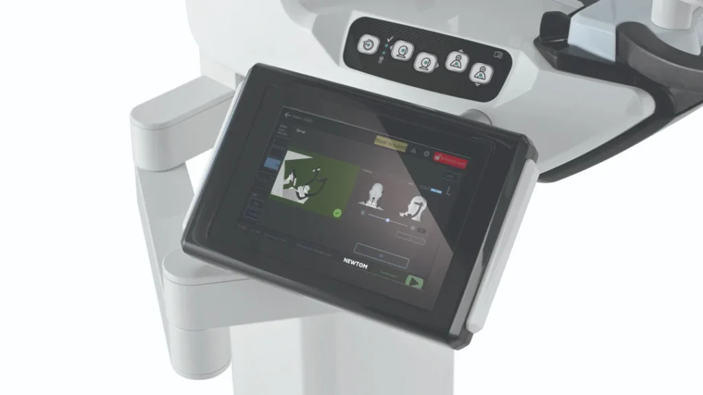
Touch screen control panel
The user-friendly interface allows you to quickly and easily manage each positioning and scanning step, ensuring a smooth workflow.
Virtual control panel
The panel can be accessed via a computer and allows doctors to monitor every stage of the examination – from selecting a diagnostic protocol to starting the scan.
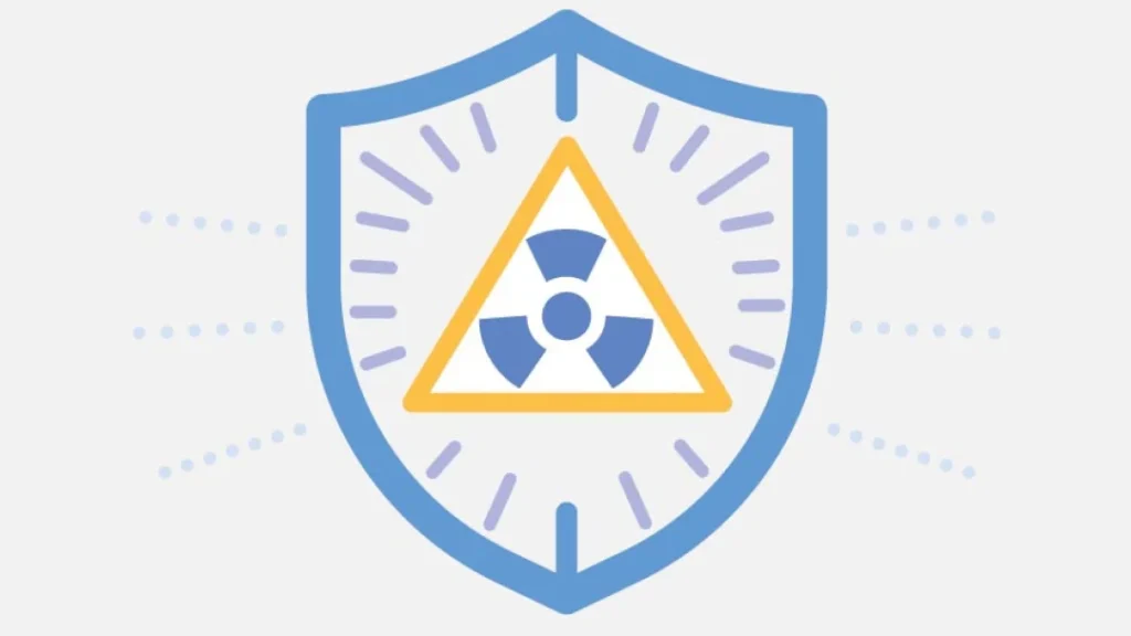
Focus on patient health
NewTom VG One prioritizes patient health. Advanced solutions ensure high-quality diagnostic images with always optimized radiation doses.
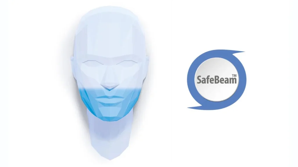
SAFEBEAM™ (patented)
This advanced technology automatically adjusts the X-ray dose before exposure and calibrates it to the patient’s physical and anatomical characteristics. This prevents unnecessary exposure and ensures detailed, consistent images without any manual tasks for the operator.
ECO protocols
These protocols, designed for 2D and 3D scanning, provide accurate diagnostic results with significantly reduced radiation dose. These modes are suitable for situations where exposure must be limited (e.g., post-operative check-ups or routine scans).
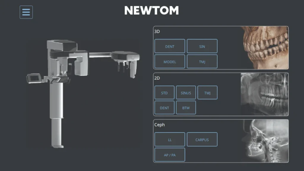
Software

One interface

Simple and convenient to use
Areas of application
The software adapts to today's growing digital demands, integrating customizations and useful features to satisfy different users ( from private clinics to radiology centers, hospitals and universities) and the needs of specialties.
- Conservative surgery
- Facial and maxillofacial surgery
- Prosthetic surgery
- Endodontics
- Gnatology
- Implantology
- Cosmetic dentistry
- Prosthetic dentistry
- Orthodontics
- Periodontology
- Radiology
2D viewer
Allows simultaneous viewing and comparison of multiple 2D and 3D images of any type compatible with the viewer. This simplifies the comparison of clinical information and increases diagnostic capabilities.
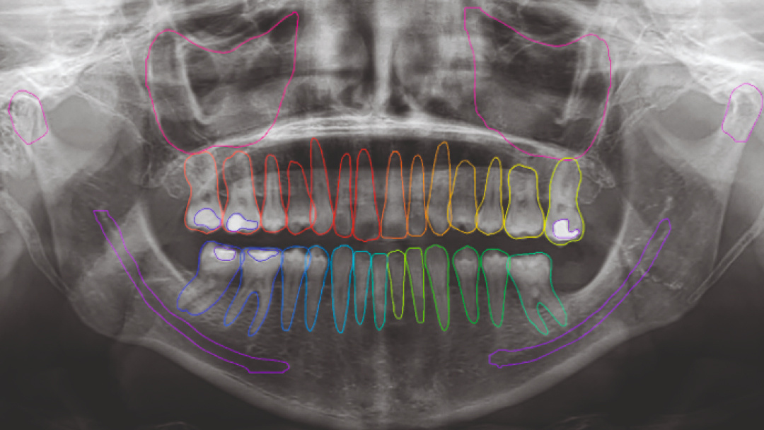
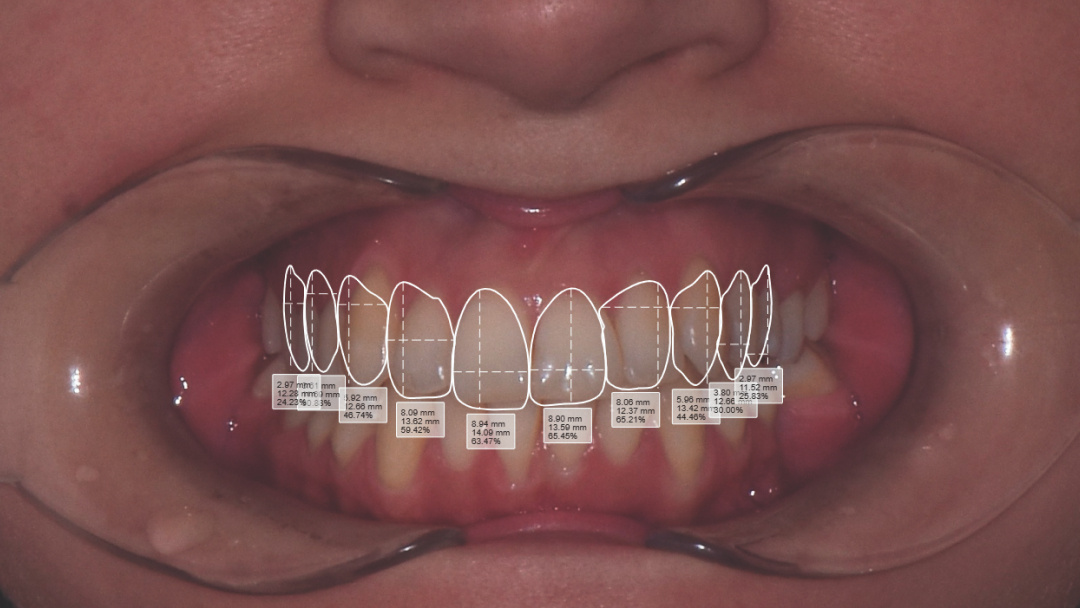
Powerful artificial intelligence tools, such as patented anatomical and pathological segmentation in both panoramic images and intraoral X-rays, provide valuable assistance for clinical analyses.
3D viewer
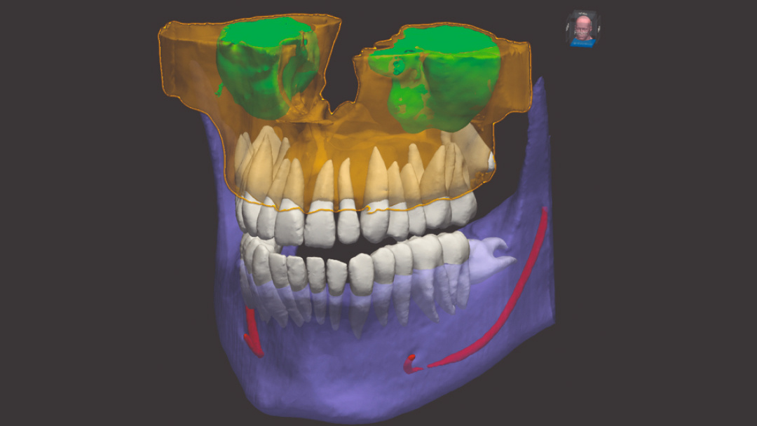
Comprehensive 3D data fusion that allows for simultaneous display of CBCT, facial scans and intraoral scans. Specialized views for endodontics, implantology and temporomandibular joint analysis.
The AI-powered clinic optimizes workflow by offering features that allow for mandibular nerve and panoramic arch tracking, automatic alignment of intraoral scans with CBCT, and segmentation of anatomical elements in CBCT.
Specific modules
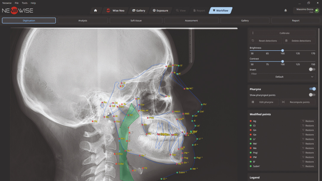
Cephalometry module
The cephalometric module uses artificial intelligence to automatically identify cephalometric points and perform a detailed analysis in seconds.

Reviews
There are no reviews yet.